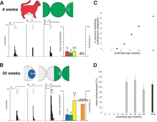
Figure 3.
Multisensory integration is absent in the earliest AES neurons and appears and matures during postnatal life. A, Shown at the top are the receptive fields (shading) and stimulus locations (icons) used in sensory testing of this auditory–somatosensory AES neuron in an 8-week-old animal. Rasters and peristimulus time histograms show the neuron responses to somatosensory (S), auditory (A), and combined (SA) stimulation. Bar graph at the right summarizes these responses and shows the lack of any multisensory interaction. Scale bar at the bottom of the histograms represents 100 ms. B, Multisensory integration in a visual–auditory neuron at 20 weeks of age. A, Auditory stimulation; V, visual stimulation; VA, combined visual–auditory stimulation. Conventions are the same as in A. *p < 0.01, t test. C, Growth in the integrating multisensory population as a function of age. D, As soon as it appears (i.e., at 12 weeks), multisensory integration is of similar magnitude to the adult. Error bars show the SDs.

