
Figure 1.
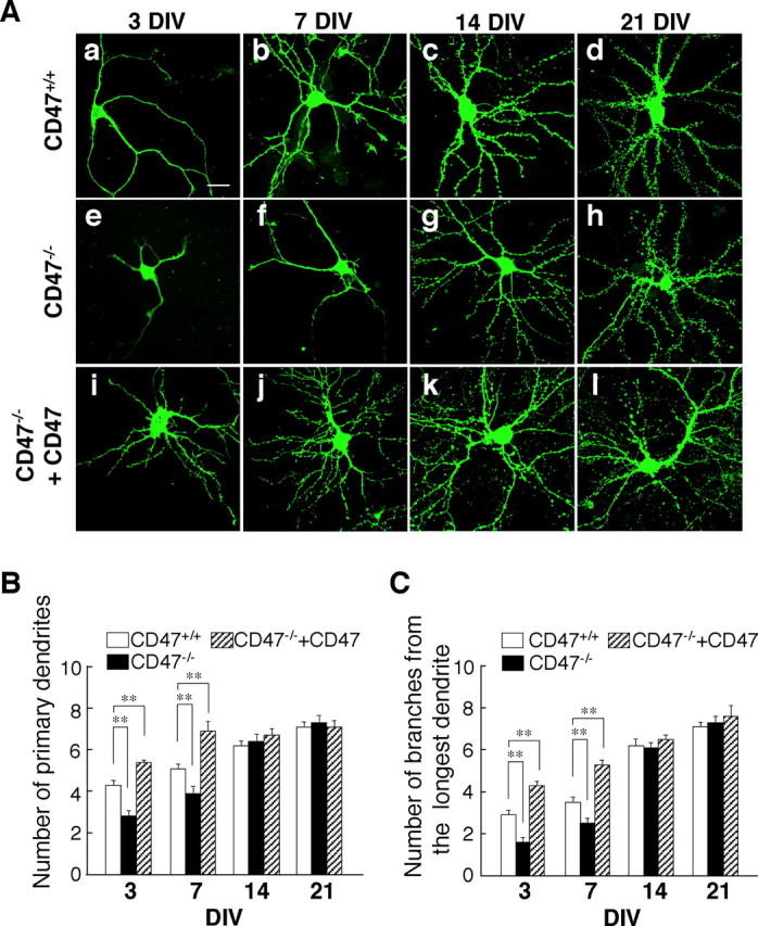
Impairment of dendritic development in cultured hippocampal neurons from CD47−/− mice. A, Hippocampal neurons isolated from newborn WT (CD47+/+) (a–d) or CD47−/− (e–l) mice were transfected with an expression vector for GFP–actin and either an expression vector for CD47 (i–l) or the corresponding empty vector (a–h) at 1 DIV (a, e, i) or 4 DIV (b–d, f–h, j–l). Neurons were fixed at 3 DIV (a, e, i), 7 DIV (b, f, j), 14 DIV (c, g, k), or 21 DIV (d, h, l), and the morphology of each neuron as revealed by GFP–actin was examined by fluorescence microscopy. Scale bar, 20 μm. B, C, Neurons treated as in A were evaluated for their dendritic morphology at the indicated times. The numbers of primary dendrites per neuron (B) and of branches from the longest primary dendrite of each neuron (C) were determined for CD47+/+ neurons (open columns), CD47−/− neurons (filled columns), and CD47−/− neurons expressing ectopic CD47 (hatched columns). Data are means ± SE of values obtained from a total of 28–36 neurons in three independent experiments. **p < 0.01 (Student's t test) for the indicated comparisons.