
Figure 4.
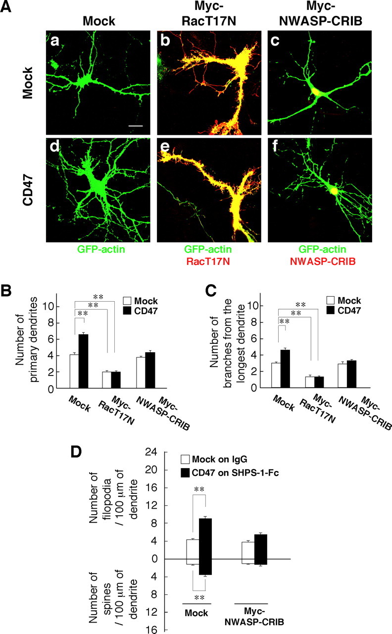
Participation of Rac and Cdc42 in the promotion of dendritic development by forced expression of CD47. A, Hippocampal neurons from WT mice were transfected at 4 DIV with an expression vector for GFP–actin, a CD47 vector (d–f) or the corresponding empty vector (a–c), and either a vector for Myc epitope-tagged RacT17N (b, e) or Myc–NWASP–CRIB (c, f) or the corresponding empty vector (a, d). At 7 DIV, the neurons were fixed and stained with an mAb to Myc (red), and GFP–actin fluorescence was also monitored (green). Neurons were also stained with an mAb to CD47 to confirm its overexpression (data not shown; see supplemental Fig. 3, available at www.jneurosci.org as supplemental material). Scale bar, 20 μm. B, C, The number of primary dendrites per neuron (B) and that of branches from the longest primary dendrite of each neuron (C) were determined for neurons treated as in A. Data are means ± SE of values obtained from a total of 30 neurons in three independent experiments. **p < 0.01 (Student's t test). D, Hippocampal neurons from WT mice were plated on dishes coated with SHPS-1–Fc or control human IgG and were transfected at 4 DIV with a vector for GFP–actin, a vector for CD47 or the corresponding empty vector, and a vector for Myc epitope-tagged NWASP–CRIB or the corresponding empty vector. At 7 DIV, the neurons were fixed and stained with an mAb to Myc, and the densities of dendritic filopodia and spines were determined. Data are means ± SE of values obtained from a total of 30 neurons in three independent experiments. **p < 0.01 (Student's t test).