
Figure 6.
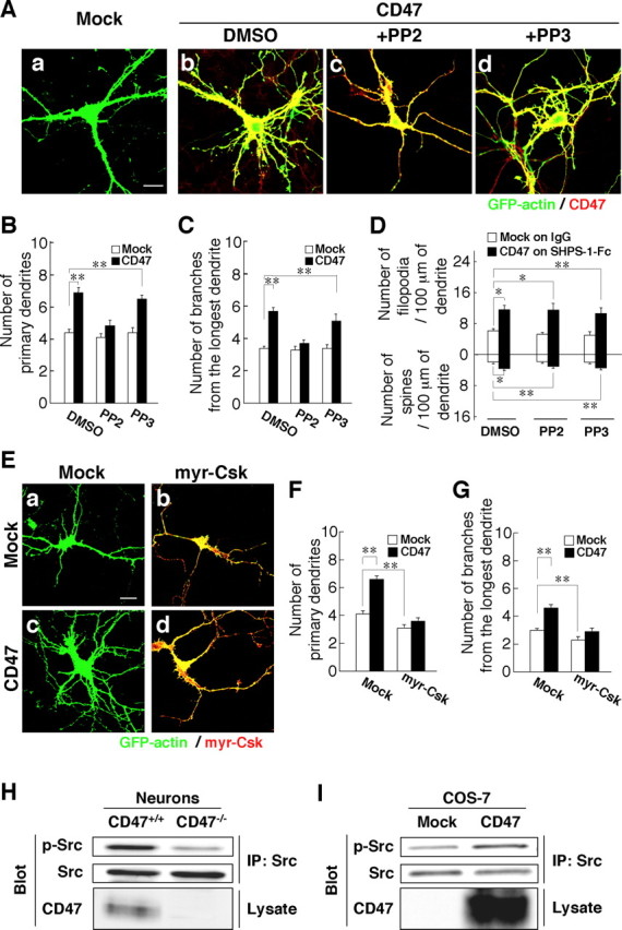
Participation of an Src family kinase in the promotion of dendritic development by CD47. A, Hippocampal neurons from WT mice were transfected at 4 DIV with an expression vector for GFP–actin and either a vector for CD47 (b–d) or the corresponding empty vector (a). They were then cultured in the presence of 20 μm PP2 (c), 20 μm PP3 (d), or dimethylsulfoxide (DMSO) vehicle (a, b). At 7 DIV, neurons were fixed and stained with an mAb to CD47 (red), and GFP–actin fluorescence was monitored (green). Scale bar, 20 μm. B–D, The number of primary dendrites per neuron (B), that of branches from the longest primary dendrite of each neuron (C), and the density of dendritic filopodia or spines (D) were determined for neurons transfected and treated with PP2 or PP3 as in A. In D, neurons transfected with the CD47 vector were plated on SHPS-1–Fc, and those transfected with the corresponding empty vector were plated on human IgG. Data are means ± SE of values obtained from a total of 33–36 neurons (B, C) or 28–32 neurons (D) in three independent experiments. *p < 0.05, **p < 0.01 (Student's t test). E, Hippocampal neurons from WT mice were transfected at 4 DIV with an expression vector for GFP–actin, with either a vector for myr–Csk (b, d) or the corresponding empty vector (a, c), and with either a vector for CD47 (c, d) or the corresponding empty vector (a, b). At 7 DIV, neurons were fixed and stained with pAbs to Csk (red), and GFP–actin fluorescence was monitored (green). Neurons were also stained with an mAb to CD47 to confirm its expression (data not shown). Scale bar, 20 μm. F, G, The number of primary dendrites per neuron (F) and that of branches from the longest primary dendrite of each neuron (G) were determined for neurons treated as in E. Data are means ± SE of values obtained from a total of 30 neurons in three independent experiments. **p < 0.01 (Student's t test). H, Cultured hippocampal neurons from CD47+/+ or CD47−/− mice were lysed at 7 DIV and subjected to immunoprecipitation (IP) with an mAb to c-Src. The resulting precipitates were subjected to immunoblot analysis both with pAbs to the Tyr416-phosphorylated form of c-Src (p-Src) and with the mAb to c-Src. Cell lysates were also directly subjected to immunoblot analysis with pAbs to CD47. Data are representative of three independent experiments. I, COS-7 cells were transfected with a vector for CD47 or the corresponding empty vector. Twenty-four hours after transfection, the cells were lysed and subjected to immunoprecipitation with an mAb to c-Src. The resulting precipitates as well as the original cell lysates were then subjected to immunoblot analysis as in H. Data are representative of three independent experiments.