
Figure 1.
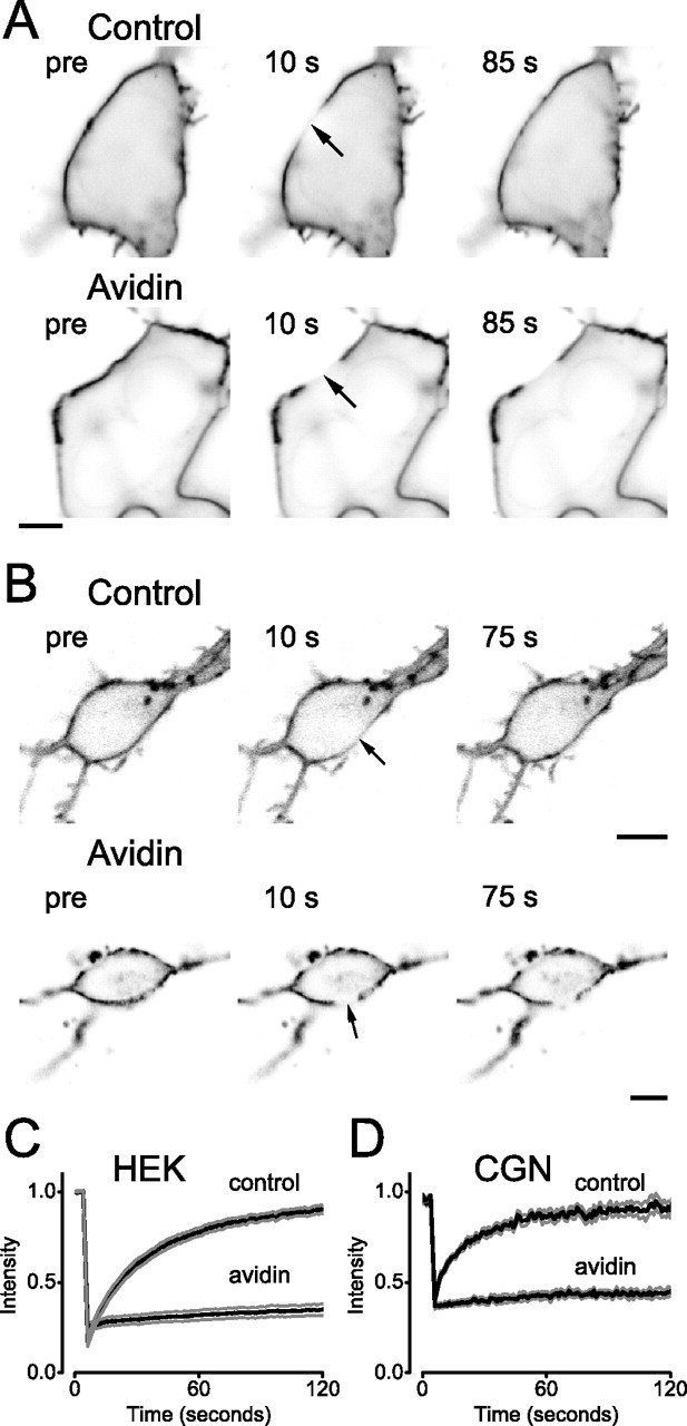
Immobilization of pH-MOR by avidin-mediated cross-linking. A, B, Confocal images of HEK 293 cells (A) or CGNs (B) expressing pH-MOR after cell surface biotinylation (Control) or cell surface biotinylation and avidin cross-linking (Avidin). Cells are shown before and at the indicated times, after photobleaching of a 5-μm-diameter ROI (arrow) centered on the plasma membrane. Scale bars, 5 μm. C, D, Fluorescence intensity in the bleached ROI is plotted versus time during FRAP experiments on control and avidin cross-linked HEK cells (C) and CGNs (D). The black traces represent the mean fluorescence intensity from 10 cells, and the gray traces represent the mean ± SEM.