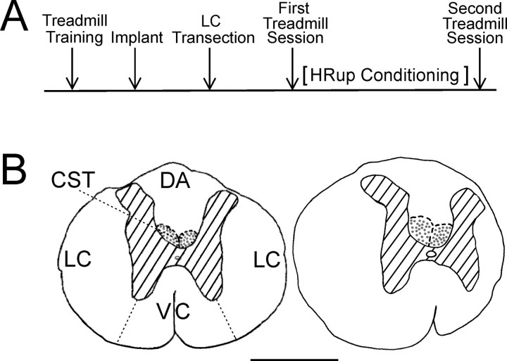
Figure 1.
A, Experimental sequence. After learning to walk on the treadmill, each rat was implanted with soleus EMG electrodes and nerve-cuff stimulating electrodes. Several weeks later, it was subjected to an LC transection. After the BBB score (Basso et al., 1995) had returned to 20–21, the first treadmill session assessed locomotion and locomotor H-reflexes. Then, the rats were (HRup rats) or were not (control rats) exposed to the HRup conditioning protocol. Finally, the second treadmill session reassessed locomotion and locomotor H-reflexes. B, Camera lucida drawings of transverse sections of T8–T9 spinal cord. Left, A normal rat with LC, dorsal column CST, dorsal column ascending tract (DA), and ventral column (VC) labeled. Right, An LC-transected rat. In the LC rat, the section shown is at the lesion epicenter. Hatching indicates gray matter, and stippled areas are the main corticospinal tract. Scale bar, 1 mm. Most of the right LC is gone.

