Figure 7.

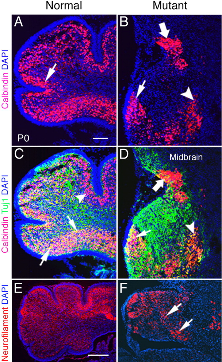
Maturation and migration of Purkinje cells are abnormal in P0 Bmpr1a;Bmpr1b knock-outs. A, At P0, most calbindin-positive Purkinje cells are localized beneath the EGL and form a distinct layer in normal animals (arrow). B, However, in mutants, most of the Purkinje cells are localized ectopically in the deep cerebellum (arrowhead) and midbrain (large arrow). Only a small number of cells are localized correctly (arrow). C, D, Detection of calbindin (red) and Tuj1 (green) immunolabeling at P0. C, Double-labeled Purkinje cells are detected in the ventral side of the cerebellum in normal neonates (arrows). Purkinje cells labeled with calbindin alone are detected in the dorsal side of the cerebellum (arrowhead). D, Double-labeled cells are located ectopically in the deep cerebellum (arrowhead) and are present in decreased number in the mutant neonates. Tuj1 expression is disorganized, and some double-labeled cells are located in the midbrain of double knock-out neonates (large arrow). Only some double-labeled cells are localized correctly (small arrow). E, In normal P0 mice, the majority of cerebellar cells express neurofilament, except those located in the EGL. F, Neurofilament expression is detected in the cerebellum in plaques (arrows) with some areas lacking expression in double mutants. Scale bars: A, 100 μm; E, 250 μm.
