Figure 1.
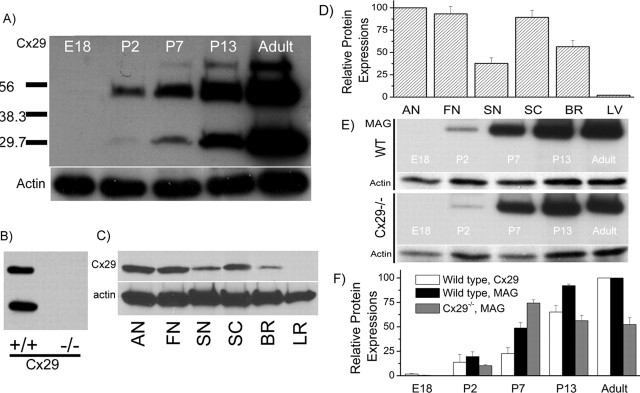
Characterization of Cx29 expression in the cochlea by Western blots. A, Western blots of Cx29 at five different developmental stages. The amount of protein loading in each lane was checked by Western blot with actin. B, Western blots of Cx29 obtained from cochlear tissues of the wild-type and Cx29−/− mice. C, Western blots comparing Cx29 expression levels in six different tissues. AN, Auditory nerve; FN, facial nerve; SN, sciatic nerve; SC, spinal cord; BR, brain (gray matter); LR, liver. D, Histogram quantifying the Cx29 protein expression level in six different tissues. E, Western blots of MAG at five different developmental stages obtained from the wild-type (top panel) and Cx29−/− (bottom panel) mice. Corresponding Western blot results with actin are given under each data panel. F, Histogram showing relative protein expression levels of Cx29 or MAG at different development stages normalized to their respective adult levels. Error bars indicate SE.

