Figure 3.

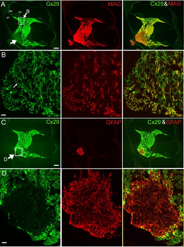
Coimmunolabeling results of Cx29 with either MAG (A, B) or GFAP (C, D). Adult mouse cochleas (2 months) were used. The images in B and D are enlarged views of the boxed areas in the left panels of A and C, respectively. A, Cochlear sections were colabeled with antibodies against Cx29 (green; left panel) and MAG (red; middle panel). A superimposed image is given in the right panel. An arrow in the left panel points to the area of weak Cx29 staining outside the glial junction in the brainstem. ST, Scala tympani; SM, scala media; SV, scala vestibule. C, Cochlear sections were colabeled with antibodies against Cx29 (green; left panel) and GFAP (red; middle panel). A superimposed image is given in the right panel. An arrow in the left panel points to the boxed area where immunoreactivities of Cx29 and GFAP around the glial juncture were compared. Scale bars: A, C, ∼400 μm; B, D, 20 μm.
