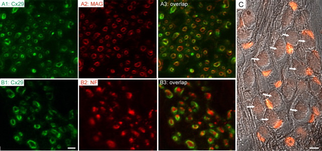
Figure 4.
Cx29 (green) coimmunolabeled with either MAG (red; A2, A3) or NF (red; B2, B3). Adult mouse cochleas (2 months) were used. Overlapped images are given in A3 and B3. C shows immunolabeling results obtained with an antibody against β-galactosidase (β-gal; red) superimposed on an image obtained with differential interference contrast optics to show the SG neurons (arrows). Scale bars: A1–A3, B1–B3, 1 μm; C, 20 μm.

