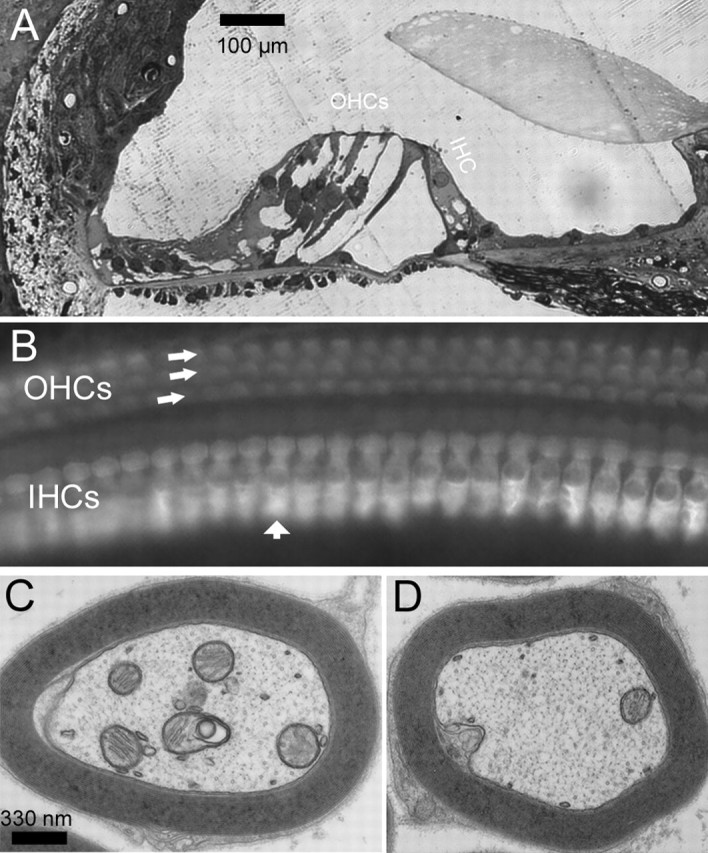
Figure 7.

Morphological examinations of hair cells and myelin around the fibers of Cx29−/− mice. A, A cochlear section showing the organ of Corti obtained from the basal region of the cochlea. B, Whole-mount view of basal inner and outer hair cells visualized by immunolabeling with an antibody against myosinVI. IHC, Inner hair cell; OHC, outer hair cell. C, D, Cross sections of myelin around auditory nerve fibers obtained from Cx29−/− (C) and wild-type (D) mice, observed with an electron microscope.
