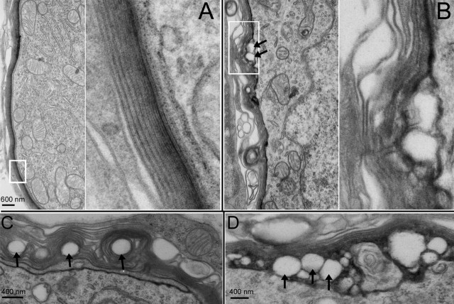
Figure 8.
Myelin morphology of wild-type and Cx29−/− mice observed with a transmission electron microscope. A, Myelin around the soma of a SG neuron obtained from a wild-type animal. The right panel presents an enlarged view of myelin outlined by a white box in the left panel. B, Myelin around the soma of a SG neuron obtained from a Cx29−/− mouse (6 months of age). The right panel presents an enlarged view of myelin outlined by a white box in the left panel. The arrows give examples of detachments between myelin and cell membrane. C, D, Examples of abnormal myelination around the soma of SG neurons observed from noise-exposed mice.

