Figure 2.
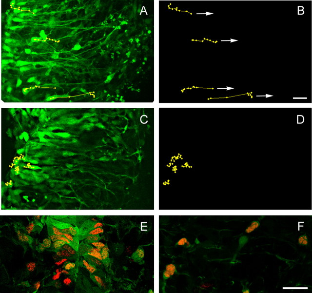
PLP-EGFP+ cells in the netrin-1 mutant spinal cord are unable to detach from the ventricular zone. A, B, PLP-EGFP+ cells in wild-type spinal cord slices show directional and radial migration toward the marginal zone as the arrows indicate (video 1, available at www.jneurosci.org as supplemental material). C, D, PLP-EGFP+ cells in mutant spinal cord slices only oscillate in the ventricular zone without translocating radially (video 2, available at www.jneurosci.org as supplemental material). Note that the overall cell morphologies of PLP-EGFP+ cells in the mutant and wild-type spinal cords are similar. In both slices, cells establish their processes toward the marginal zone. In A and C, the ventral midline is located at the left edge of the panel. E, F, PLP-EGFP+ cells at E12.5 express Olig2, a marker for OPCs. The majority of PLP-EGFP+ cells in the ventricular zone (E) also express Olig2 proteins, suggesting they represent OPCs. F, PLP-EGFP+ cells in the intermediate zone also express Olig2 with few exceptions. Scale bars: (in B) A–D, 20 μm; (in F) E, F, 25 μm.

