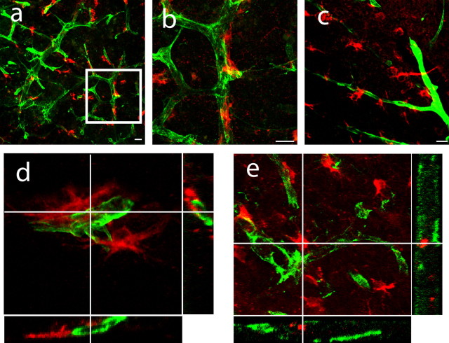
Figure 4.
Peri-infarct neuroblasts form close physical associations with vascular endothelial cells. a, b, DCX+ cells (red) form close physical associations with peri-infarct blood vessels immunoreactive for laminin (green). The region within the box in a is enlarged in b. c, PSA-NCAM+ cells (red) align with endothelial cells stained for PECAM-1 (green). Scale bar, 25 μm. d, e, Three-dimensional confocal reconstructions demonstrate DCX+ cells (red) interdigitate within the folds of laminin+ (d) and PECAM-1+ endothelial cells (e, green) in peri-infarct cortex.

