
Figure 5.
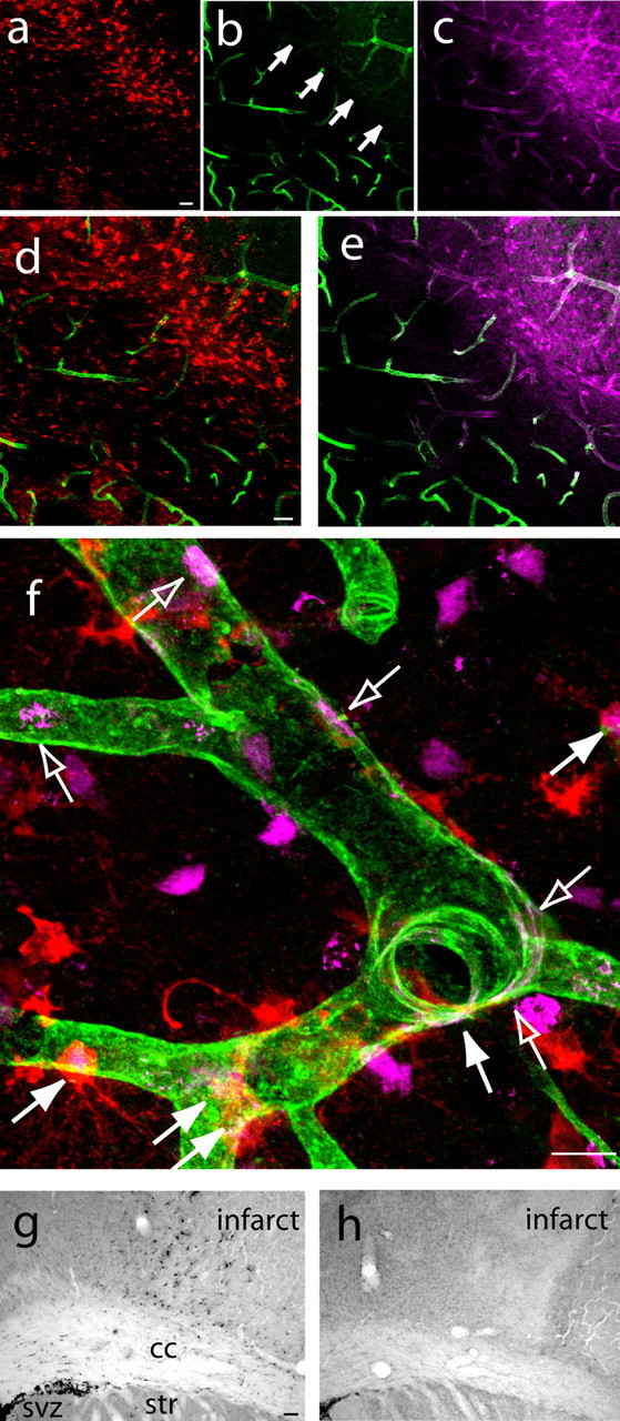
Neurogenesis and angiogenesis are causally linked in a novel neurovascular niche in peri-infarct cortex. a–e, DCX+ cells (a) localize to a region of vascular remodeling in peri-infarct cortex characterized by poor lectin perfusion (b) but robust PECAM-1 immunoreactivity (c) at day 5 after stroke. d and e represent merged images of a,b and b,c, respectively. Arrows in b indicate the region of poor lectin perfusion adjacent to the infarct core. Scale bars, 50 μm. f, High-magnification confocal image showing that newly born neuroblasts localize in a region of newly born endothelial cells at day 7 after stroke. Filled arrows indicate newly born neuroblasts double labeled for DCX (red) and BrdU (purple), and open arrows indicate newly born endothelial cells double labeled for PECAM-1 (green) and BrdU (purple). Scale bar, 25 μm. g, h, DCX+ cells, stained with DAB, are significantly reduced in peri-infarct cortex of endostatin-treated (h) compared with vehicle-stroke animals (g) at day 7 after stroke. Scale bar, 100 μm. cc, Corpus callosum; str, striatum.