
Figure 2.
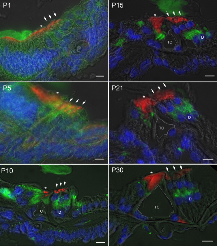
Developmental expression of prosaposin in rat cochlea. Immunofluorescence staining of prosaposin (green), rhodamine-phalloidin (red), and DAPI (blue) with a semitranslucent transmitted light micrograph overlay from P1 through P30 demonstrates initially diffuse expression of prosaposin throughout the primitive organ of Corti that gradually localizes to several discrete locations within the organ of Corti, including the IHCs and its supporting cells, inner pillar cells, and synaptic region of the OHCs (either base of OHCs or apex of the DCs). At P5 there is increased signal in the regions of the IHC and OHC relative to surrounding tissues. By P10, labeling can be clearly differentiated in the region of the basal OHCs or apical DCs, IHCs, and inner pillar cells. This labeling pattern is refined, with further loss of label in surrounding cells through P30, with localization to regions surrounding the IHC, inner pillar cell, and base of OHC and/or apex of DC. In each panel, an asterisk is placed above the IHC, three small arrows are placed above the OHC, and the central of the three Deiters' cells is marked by a D where clearly visible (after P10). The tunnel of Corti is outlined in white and labeled TC. Scale bars, 10 μm.