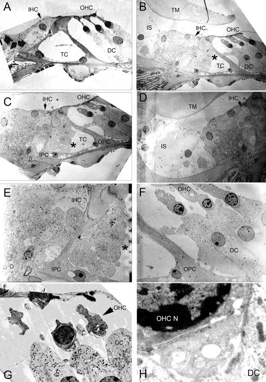
Figure 7.

Cochlear electron microscopy of the prosaposin knock-out mice. A, Wild-type organ of Corti demonstrating normal anatomy. B, C, In the prosaposin KO mouse, there is marked cellular hypertrophy in the region of the IHCs. B, C, E, There is bulging tissue into the tunnel of Corti (TC, asterisk). D, Hypertrophy of the inner sulcus (IS) region. F–H, In the region of the OHCs, DCs are hypertrophied and there is vacuolization within the OHCs. H, The base of the OHC, efferent synaptic cleft, and apex of the DC. The synaptic cleft between the OHC and the DCs is abnormally enlarged and vacuolization is seen in the cells. TM, Tectorial membrane; OPC, outer pillar cell.
