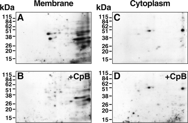
Figure 3.
Broad pH range analysis and identification of plasminogen-binding proteins that expose C-terminal lysines on the PC12 cell surface. Intact PC12 cells were incubated either with 100 U/ml CpB (B, D) or buffer (A, C) for 30 min at 37°C before fractionation into membrane or cytosolic fractions. 2D-PAGE was performed on 100 μg of membrane proteins (A, B) or 10 μg of cytoplasmic proteins (C, D), followed by ligand blotting with 125I-plasminogen. In specificity controls, the binding of 125I-plasminogen in the ligand blots was specific because no plasminogen binding spots were detected in the presence of 0.1 m EACA.

