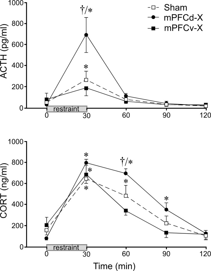
Figure 5.
Effects of mPFC lesions on pituitary–adrenal responses to acute restraint. Mean ± SEM plasma ACTH (top) and corticosterone (CORT; bottom) levels in sham-, mPFCd-, and mPFCv-lesioned animals measured before and after a single 30 min restraint session. Top, Stress exposure significantly increases plasma levels of ACTH in sham-lesioned animals (at 30 min), and mPFCd lesions produce a marked (2.6-fold) enhancement of this effect. In contrast, plasma ACTH in mPFCv-lesioned animals is not significantly elevated over pre-stress levels (p = 0.2). Bottom, Stress exposure also significantly increases plasma CORT levels in sham-lesioned animals (at 30 and 60 min). mPFCd lesions result in a prolonged increase in stress-induced plasma CORT (p < 0.05 at 30, 60, and 90 min compared with 0 min), whereas mPFCv-lesioned animals show a more rapid recovery to pre-stress levels (p < 0.05 at 30 min only). *p < 0.05, differs significantly from basal (0 min) values from within each group; †p < 0.01, differs significantly from sham-lesioned animals. n = 5 per group.

