
Figure 3.
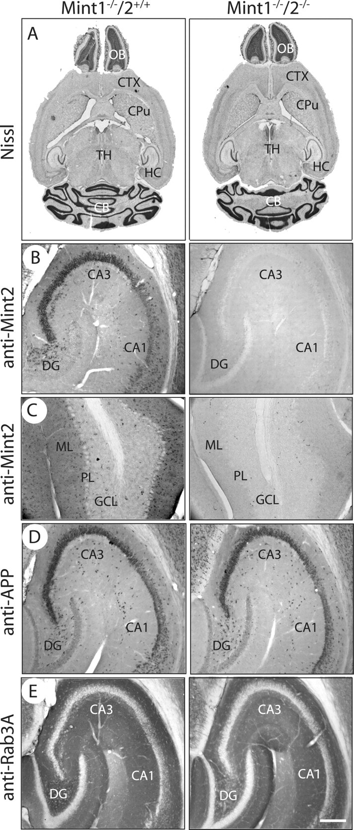
Overall brain structure and morphology of adult Mint KOs. A, Nissl brain sections of Mint 1−/−/2+/+ (left) and Mint 1−/−/2−/− double KO mice (right) at 6–8 weeks of age showed normal brain architecture. CTX, Cerebral cortex; CPu, caudate–putamen; TH, thalamus; OB, olfactory bulb; HC, hippocampus; CB, cerebellum. B, C, Mint 2 immunostaining was abolished in Mint 1−/−/2−/− double KO mice (right) as expected in the hippocampus (B) and cerebellum (C) compared with Mint 1−/−/2+/+ littermate control mice. DG, Dentate gyrus; ML, molecular layer; PL, Purkinje cell layer; GCL, granule cell layer. D, E, Immunostaining for APP (D) and Rab3A (E) displayed normal and distinct localization in the hippocampus in Mint 1−/−/2+/+ and Mint 1−/−/2−/− double KO littermate mice. Scale bar: (in E) B–E, 100 μm.