
Figure 5.
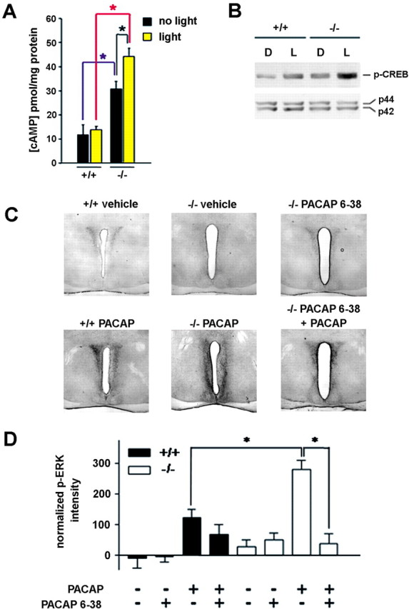
Dexras1 modulates cAMP-dependent signaling and PACAP/PAC1-mediated MAPK activation in the SCN. A, Wild-type and dexras1−/− mice received a single light pulse (5 min, 40 lux) at CT 20, and the SCN was dissected immediately and analyzed for cAMP content by ELISA. Control subjects did not receive a light pulse but were killed at the same circadian time. Light did not increase cAMP levels in wild-type mice (+/+). However, dexras1−/− mice (−/−) exhibited a significant light-induced increase in cAMP. Additionally, under basal conditions, dexras1−/− mice had significantly elevated cAMP levels relative to wild-type mice. Values are presented as mean ± SEM pmol cAMP per mg of protein. n = 4–5 per group. *p < 0.05 (two-way ANOVA). B, Western blot analysis of light-induced p-CREB expression in the SCN. Wild-type and dexras1−/− mice received a single light pulse (15 min, 40 lux) at CT 20, and the SCN was dissected 30 min after the start of the light exposure. Pooled SCN extracts were probed for the expression of p-CREB. Expression of total ERK1/2 (p42, p44) was used as the loading control. L denotes light-treated mice, whereas D denotes dark controls. C, D, Wild-type (+/+) and dexras1−/− (−/−) mice were infused with the PAC1 antagonist PACAP 6–38 (500 μm; 3 μl) via a guide cannula positioned in the lateral ventricle 30 min before a second infusion of PACAP (40 μm; 3 μl). Thirty minutes after the second infusion, mice were killed, and brain sections were processed for p-ERK. ERK activation was compared with mice that received infusions of vehicle only, PACAP alone, or PACAP 6–38 alone. Representative micrographs are provided in C. Quantitation of p-ERK expression in the SCN is given in D. Data are presented as mean ± SEM p-ERK-immunoreactive nuclei per SCN section. There was a significant effect of genotype on PACAP-mediated ERK activation as well as a significant effect of PACAP 6–38 on PACAP-induced p-ERK expression in the knock-out SCN. n = 6–7 per group. *p < 0.01 (two-tailed Student's t test).