
Figure 7.
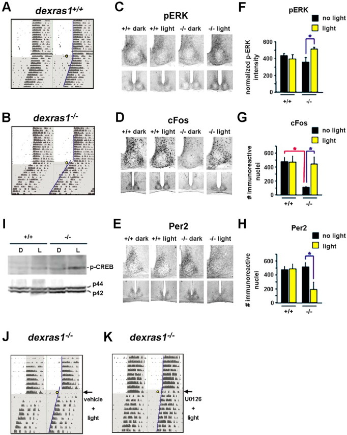
The absence of Dexras1 reveals a mid-day photic response in the SCN. A, B, Representative actograms of wheel-running activity of dexras1+/+ (A) and dexras1−/− (B) mice. Mice were exposed to a single 40 lux light pulse for 15 min at ZT 8 (yellow circle), exactly as described in Figure 1. Activity onsets are indicated by blue lines. C–E, Immunohistochemical analysis of p-ERK (C), c-Fos (D), and Per2 (E) expression in response to a single 5 min (for p-ERK) or 15 min (for c-Fos and Per2) light pulse at CT 6. Wild-type (+/+) and knock-out (−/−) mice were killed immediately (for p-ERK), 2 h (for c-Fos), or 6 h (for Per2) after the light treatment. Dark control animals were not exposed to light but were killed at the same circadian times. Light exposure in the subjective daytime had no effect on p-ERK, c-Fos, and Per2 levels in the SCN of wild-type mice. Baseline levels of c-Fos were reduced in dexras1−/− mice relative to wild-type controls. A single light pulse in the mid-subjective day increased p-ERK and c-Fos immunoreactivity in the SCN of dexras1−/− mice. Per2 immunoreactivity was decreased in dexras1−/− SCN after light treatment. F–H, Quantitation of p-ERK (F), c-Fos (G), and Per2 (H) expression in the SCN. Data are presented as mean ± SEM densitometric intensity (for p-ERK) or mean ± SEM number of immunoreactive nuclei per SCN section (for c-Fos and Per2). n = 4–6 per group. *p < 0.05 (two-tailed Student's t test). I, Western blot analysis of light-induced p-CREB expression in the SCN. Wild-type (+/+) and dexras1−/− (−/−) mice received a single light pulse (15 min, 40 lux) at CT 6, and the SCN was dissected 30 min after the start of the light exposure. Pooled SCN extracts were probed for the expression of p-CREB. Expression of total ERK1/2 (p42, p44) was used as the loading control. L denotes light-treated mice, whereas D denotes dark controls. J, K, U0126 inhibits ZT 8 light-induced phase advances in dexras1−/− mice. Representative actograms of wheel-running activity of dexras1−/− mice are shown. Mice were entrained to a 12 h LD schedule (400 lux). On the day of the experiment, lights remained off at ZT 0. Thirty minutes before a 15 min light pulse of 40 lux at ZT 8 (yellow circle), mice were infused with vehicle (DMSO) (J) or U0126 (10 mm; 3 μl) (K) via a guide cannula placed in the third ventricle. Subsequent to the light pulse, animals were maintained in DD for at least 7 d. Activity onsets are indicated by blue lines.