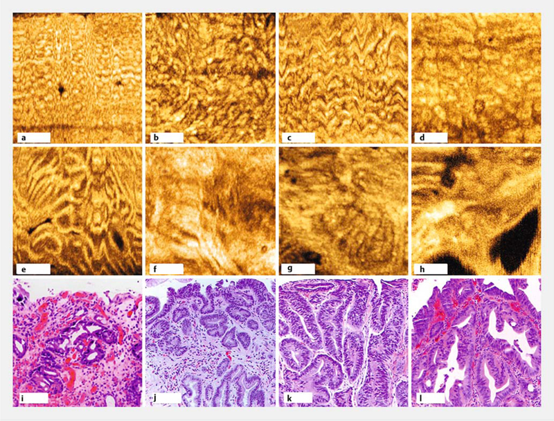
►Fig.3.

En face optical coherence tomography (OCT) features. a-d Representative regular mucosal patterns from nondysplastic Barrett’s esophagus showing size and shape variations. e-h Irregular mucosal patterns from neoplasia showing branching, distortion, and absence of mucosal patterns. i-l Hematoxylin and eosin histology indicating low grade dysplasia (i), high grade dysplasia (j and k), and esophageal adenocarcinoma (l) correlated with datasets e-h, respectively. En face OCT cropped to highlight regions of interest. Scale bars 500 μm (a-h), 100μm (i-l).
