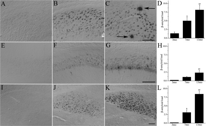
Figure 1.
Aβ accumulation increases with age in male 3xTg-AD mice. Shown are representative photomicrographs of Aβ immunoreactivity in sham GDX 3xTg-AD mice at ages 3 (A, E, I), 7 (B, F, J), and 13 (C, G, K) months in subiculum (A–C), hippocampus CA1 (E–G), and amygdala (I–K). Arrows show extracellular Aβ deposits. Scale bars, 100 μm. Aβ immunoreactivity in 3-, 7-, and 13-month-old 3xTg-AD mice was quantified by load values in subiculum (D), CA1 (H), and amygdala (L). Data show the mean load values ± SEM. *p < 0.05 versus 3 month group; # p < 0.05 versus 7 month group.

