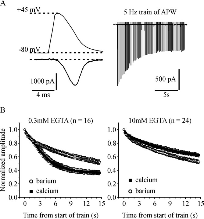
Figure 1.
Both voltage-dependent and Ca2+-dependent inactivation of ICa play a significant role during a sustained train of APW. A, The top left shows the APW used to stimulate the cells, and the bottom left shows a representative ICa. The right panel shows a series of ICa elicited by a train of APW applied at 5 Hz for 15 s. B, Cells were stimulated with a 5 Hz train of APW first in extracellular solution containing 5 mm Ca2+ and then in solution containing 5 mm Ba2+. Peak current amplitude elicited by each APW was normalized to the first pulse within the train and plotted against time. The left panel shows data from cells recorded with 0.3 mm EGTA in the patch-pipette solution. The decline in current amplitude during the train was significantly greater in Ca2+ than in Ba2+ (p < 0.0001) indicating that Ca2+-dependent inactivation plays a role. The right panel is from cells recorded with 10 mm EGTA in the patch-pipette solution. The decline in current amplitude during the train was slightly but significantly less in Ca2+ than in Ba2+ (p < 0.002). Error bars indicate SEM.

