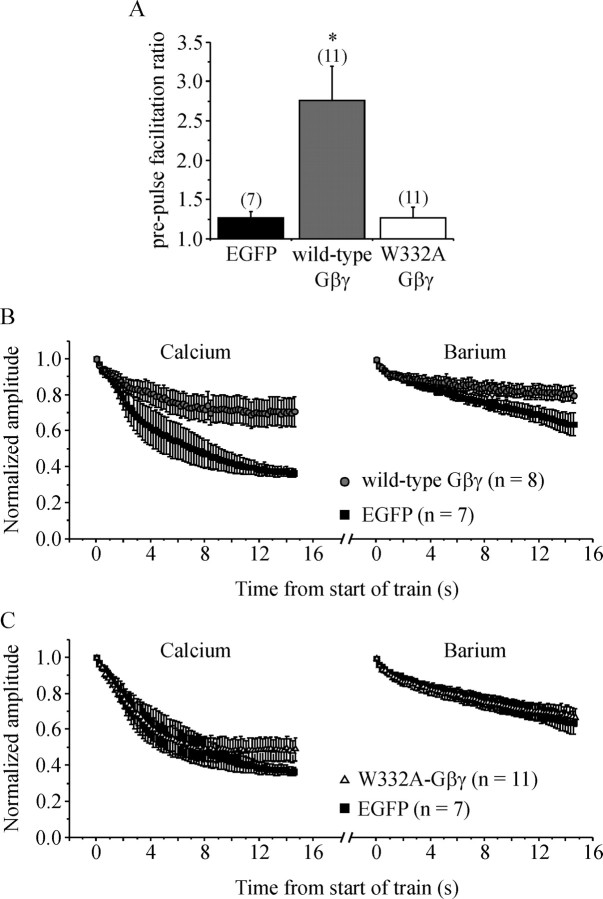
Figure 3.
G-protein βγ subunits reduce inactivation of ICa. Cells were transiently transfected with Gγ2 and either wild-type Gβ1 or a point mutant (W332A) of Gβ1. The Gβ subunits were tagged with EGFP to enable visual identification of transfected cells. Control cells were transiently transfected with EGFP. A, Mean prepulse facilitation of ICa produced by a conditioning step to +100 mV is plotted. Cells transfected with wild-type Gβγ showed strong prepulse facilitation of ICa (*p < 0.001 compared with controls) because of reversal of tonic inhibition of ICa. In cells transfected with W332A-Gβγ, facilitation was not significantly different from control (EGFP) cells, confirming that W332A-Gβγ does not bind to and inhibit Ca-channels. B, Control cells and cells expressing wild-type Gβγ were stimulated with a 5 Hz train of APW in Ca2+-containing and then in Ba2+-containing recording solutions. Current amplitude was normalized to the first APW of the train and plotted against time. Wild-type Gβγ significantly reduced inactivation compared with control cells in both Ca2+ (p < 0.003) and Ba2+ (p < 0.04) recording solutions. C, Same layout as B but comparing control cells and cells expressing W332A-Gβγ. Inactivation was not significantly reduced by W332A-Gβγ. Error bars indicate SEM.

