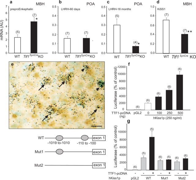
Figure 5.
Hypothalamic expression of TTF1 target genes is altered in 60-d-old Ttf1SynCre KO mice. a, Preproenkephalin mRNA levels are increased in the MBH. Normally, TTF1 represses preproenkephalin gene transcription. b, LHRH mRNA abundance in the POA is similar to WT mice at 60 d. c, However, the age-related increase in LHRH mRNA levels seen in WT mice is abolished in Ttf1SynCre KO animals. Normally, TTF1 transactivates the LHRH promoter. d, KiSS1 mRNA abundance in the MBH is decreased. e, KiSS1 neurons of the arcuate nucleus identified by in situ hybridization (black grains) also express TTF1 protein, identified by immunostaining (brown color). Examples of colocalization are denoted by arrows. Some KiSS1 mRNA-containing cells are TTF1 negative (arrowheads). Scale bar, 20 μm. f, TTF1 transactivates the KiSS1 promoter, as assessed by functional promoter assays using a luciferase reporter gene. g, Deletion of either a single proximal putative TTF1 recognition motif [located at −110 to −100 relative to the presumed transcription start site; mutation 1 (Mut1)] or both this motif and an additional site (located at −1019 to −1010; Mut2) in the 5′ flanking region of the hKiSS1 gene obliterates the transactivating effect of TTF1 on the KiSS1 promoter. *p < 0.05; ***p < 0.001.

