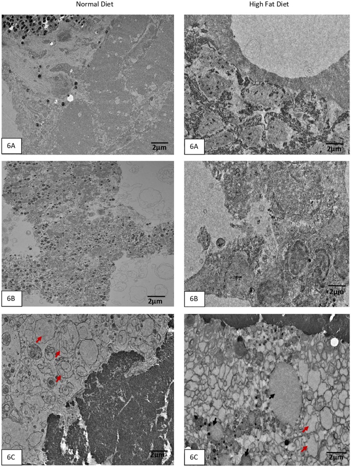
Fig 6.
A-C. Transmission Electron Microscopy of zebrafish liver sections at medium magnification show the presence of neutral fat with HFD in normal Tab-5 Fig 6A, transgenic with normal GCKR Fig 6B, and transgenic with mutant GCKR Fig 6C. Neutral fat appears as small round vesicles with no structure. There is abnormal fat accumulation in the transgenic mutant fish with HFD (Fig 6C). Black arrows point to round vesicle like structures with empty content which are fat droplets. The red arrows point to two huge abnormal structures which appear like fusion of large neutral fat vesicles in the transgenic mutant with HFD. However, the transgenic mutant with normal diet shows abnormal structures with possible accumulation of phospholipids (red arrows) in hepatic cells suggesting abnormal metabolic function.

