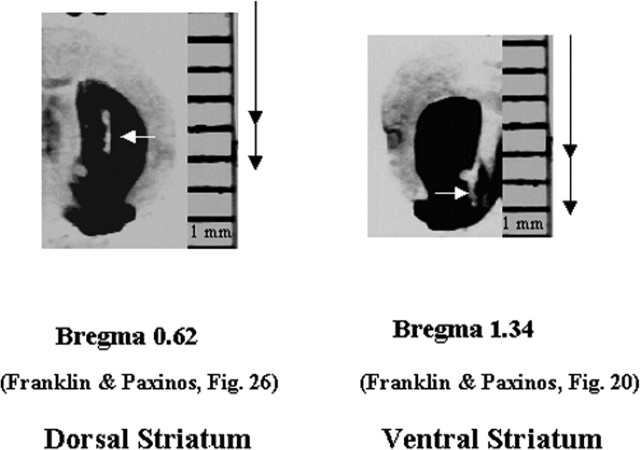
Figure 1.
Placement of the dialysis probes. The micrograph in the left panel shows the probe placement in the dorsal striatum of a representative mouse in this study. The micrograph in the right panel shows the probe placement in the ventral striatum from a representative mouse in this study. Arrows indicate the track of the microdialysis probe, which extends 2 mm below the guide cannula. To the right of each figure, the top arrows indicate the extent of the guide cannula, and the bottom arrows approximate the microdialysis probe extending 2 mm beyond the guide cannula.

