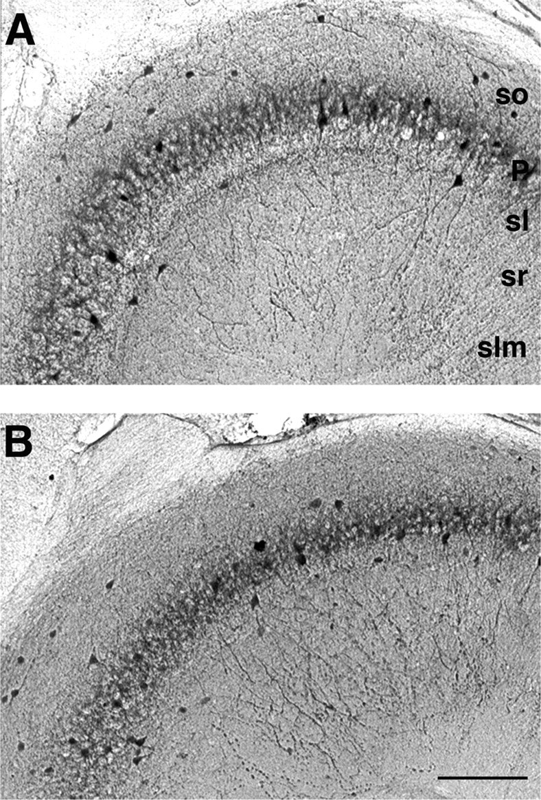
Figure 6.

Numbers of parvalbumin-immunopositive interneurons are unchanged in area CA3 from LPA1−/− mice. The distribution of parvalbumin-immunoreactive neurons in area CA3 of WT (A) and KO (B) mice is shown. Parvalbumin-immunopositive cells were counted in microscopic view fields, as shown in example panels of CA3 region. No significant changes in the number of parvalbumin-immunopositive neurons were observed between WT (A) and KO (B) hippocampus (see Results). so, Stratum oriens; P, stratum pyramidale; sl, stratum lucidum; sr, stratum radiatum; slm, stratum lacunosum-moleculare. Scale bar: (in B) A, B, 140 μm.
