
Figure 3.
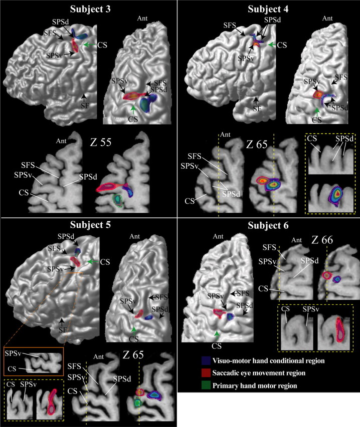
The location of the premotor hand region for conditional motor responses is in blue, the location of the saccadic eye movement region (i.e., the FEF) is in red, and the location of the primary motor cortex hand representation is in green in subjects 3–6. The foci of activity illustrated result from the subtractions reported in Tables 1–3. These foci are shown on each subject’s left hemisphere: lateral view (top left diagram) and top view (top right diagram). The green arrow indicates the point of the central sulcus in the depth of which the primary hand motor representation is located (i.e., Broca’s pli de passage moyen). For each subject, horizontal sections at different levels (z coordinate) in standard stereotaxic space are shown. The left horizontal sections are anatomical MRIs, and the right sections are the same ones with the premotor hand conditional (blue) and saccadic eye movement (red) foci displayed. In subjects 3 and 5, the primary hand motor region is also displayed in green. In subjects 4–6, the yellow dotted line indicates the level of the sagittal section (i.e., the x coordinate) illustrated within the yellow dotted box. Note that, in subject 5, the precentral gyrus has receded and therefore the SPSv and the central sulcus blend in certain locations. This can be appreciated by careful inspection of the horizontal section indicated by the orange line and box. Ant, Anterior part of the brain; CS, central sulcus; SF, Sylvian fissure.