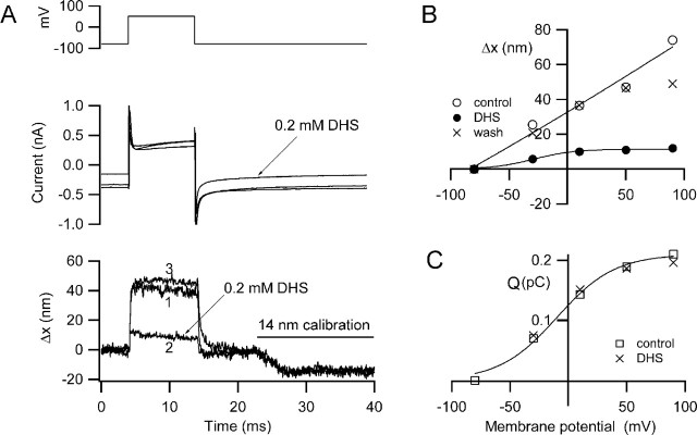
Figure 2.
Depolarization-induced hair bundle motion and its block by dihydrostreptomycin. A, Membrane currents (middle) and hair bundle movements, Δx, (bottom) for depolarization to +50 mV before (1), during (2), and after (3) recovery from application of 0.2 mm DHS. DHS reduces the maximum movement and also blocks part of the current turned on at the holding potential (−84 mV). For all Δx traces, movement toward the tip of the V is denoted as positive, and the negative deflections at the end of the traces is a 14 nm calibration signal. B, Voltage dependence of the movements (Δx) before, during, and after DHS application. C, Voltage dependence of charge movement (Q) before and during DHS perfusion. The charge movement (Q), calculated from the current transients at the onset of the depolarizing step, reflects activation of the prestin motor and is unaffected by DHS. Bathing solution contains 0.1 mm Ca2+ and 1 mm Mg2+. P9 rat, CsCl intracellular, maximum MET current of 0.77 nA.

