
Figure 3.
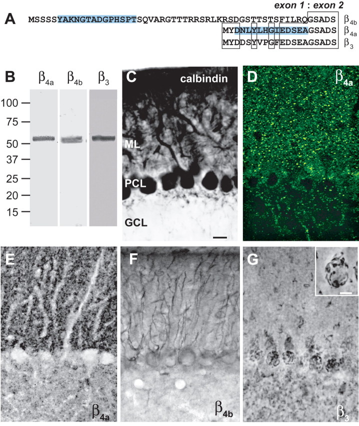
Distribution of β subunit subtypes in mouse cerebellum. A, Splice variant-specific antibodies were created against unique β4a and β4b sequences (highlighted). B, Western blot showing that β4a-, β4b-, and β3-specific antibodies recognize 52–55 kDa proteins in sucrose step gradient-purified mouse cerebellar membranes. Numbers to the left indicate molecular mass in kilodaltons. C, Purkinje cells labeled with anti-calbindin antibody (scale bar, 20 μm) serve to distinguish the molecular (ML), Purkinje cell (PCL), and granule cell (GCL) layers (for orientation in D–G). D, E, Punctate labeling of the β4a subunit in the molecular layer of the cerebellum: D, confocal microscopy, 1-μm-thick image; E, light microscopy, 50-μm-thick section. F, The β4b subunit is expressed in Purkinje cell bodies and Bergman glia. G, The β3 subunit is expressed in basket cell structures surrounding Purkinje cell bodies and in granule cell layer puncta. Inset, Higher-power image (scale bar, 10 μm) of basket cell labeling (supplemental Fig. 2, available at www.jneurosci.org as supplemental material).