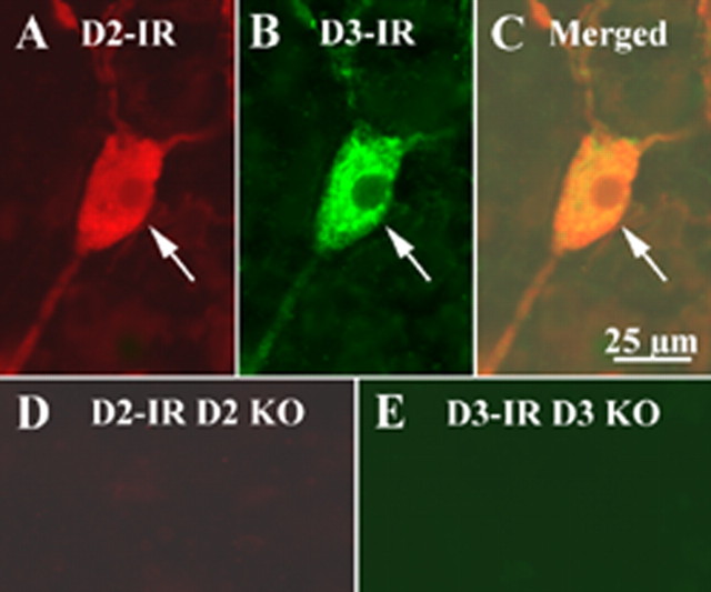
Figure 6.
D2 and D3 immunocytochemistry were performed on the submucosal plexus of the ileum of CD-1 mice. The same tissue preparation from D2 and D3 KO mice was used as control. D2 receptor immunoreactivity (IR) was revealed by a D2 rabbit antibody and a donkey anti-rabbit Alexa 594 secondary antibody. D3 receptor immunoreactivity was revealed by a D3 goat antibody, a biotinylated donkey anti-goat secondary antibody, and streptavidin FITC. On the tissue of CD-1 mice, D2-immunoreactive products were present in the enteric neurons (A), and D3-immunoreactive products were also present in the enteric neurons (B). D2 and D3 immunoreactivities were colocalized in the same cell (C). However, D2 immunoreactivity was not detected on the tissue of D2 knock-out (KO) mice (D); no D3 immunoreactivity was detected on the tissue of D3 knock-out mouse (E). The arrows indicate the immunoreactive neurons. Scale bar: (in C) 25 μm.

