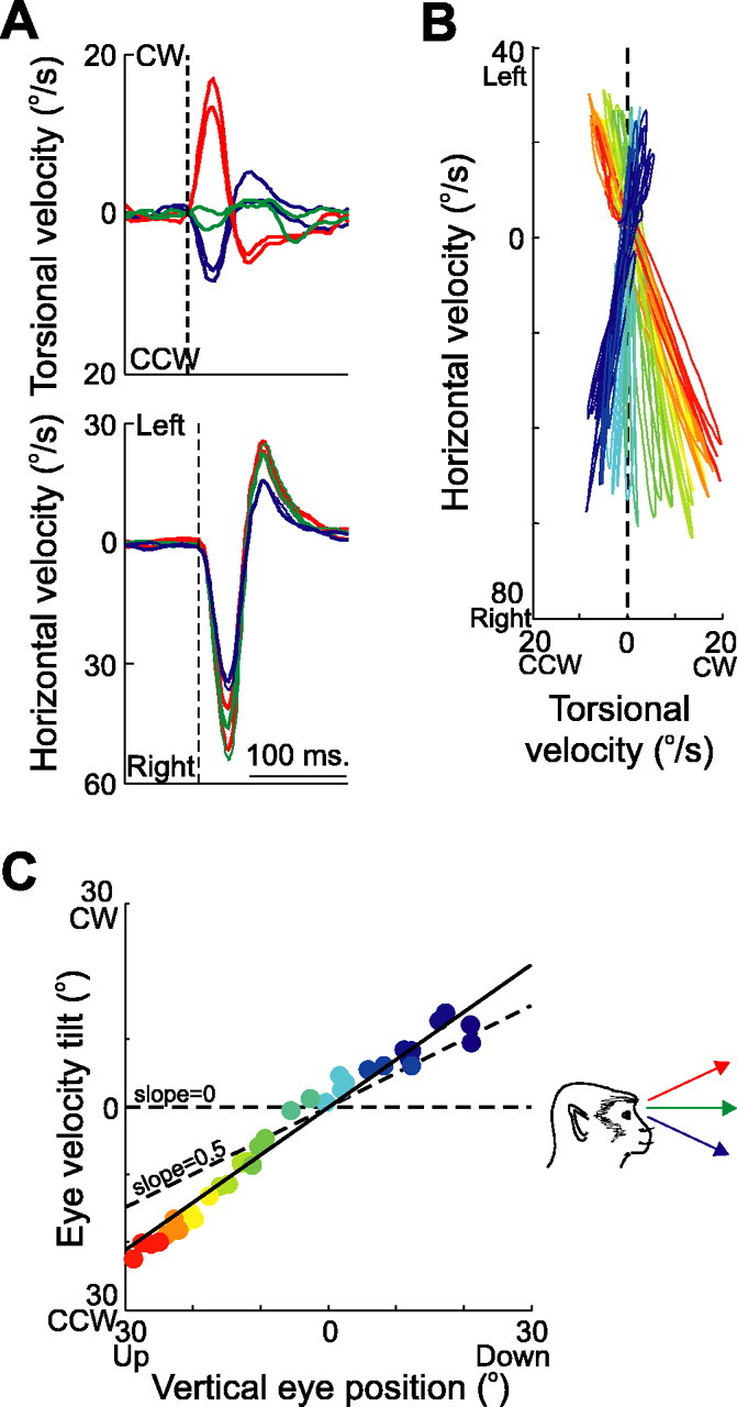
Figure 3.

Characteristics of eye velocity during abducens nerve microstimulation. A, Temporal plots of eye velocity from different initial eye positions (green, straight ahead; red, 25° up; blue, 25° down) are similar in the horizontal domain (bottom) but differ in the torsional domain (top). Two traces of each are shown. B, Horizontal versus torsional velocity is plotted as the eye assumes different vertical positions. The orientation of the eye when the stimulation was delivered is illustrated by a color palette, ranging from 25° up (dark red) to 25° down (dark blue) in 5° intervals. The orientation of Listing’s plane is indicated by the vertical dashed line at 0° torsion. C, Measures of eye velocity tilt angle are plotted against initial vertical eye position. A slope of 0 (indicating fixed axes of rotation and a zero-angle rule) and a slope of 0.5 (indicating eye position-dependent axes of rotation and proper implementation of the half-angle rule) are also shown (dashed lines). CCW, counterclockwise; CW, clockwise.
