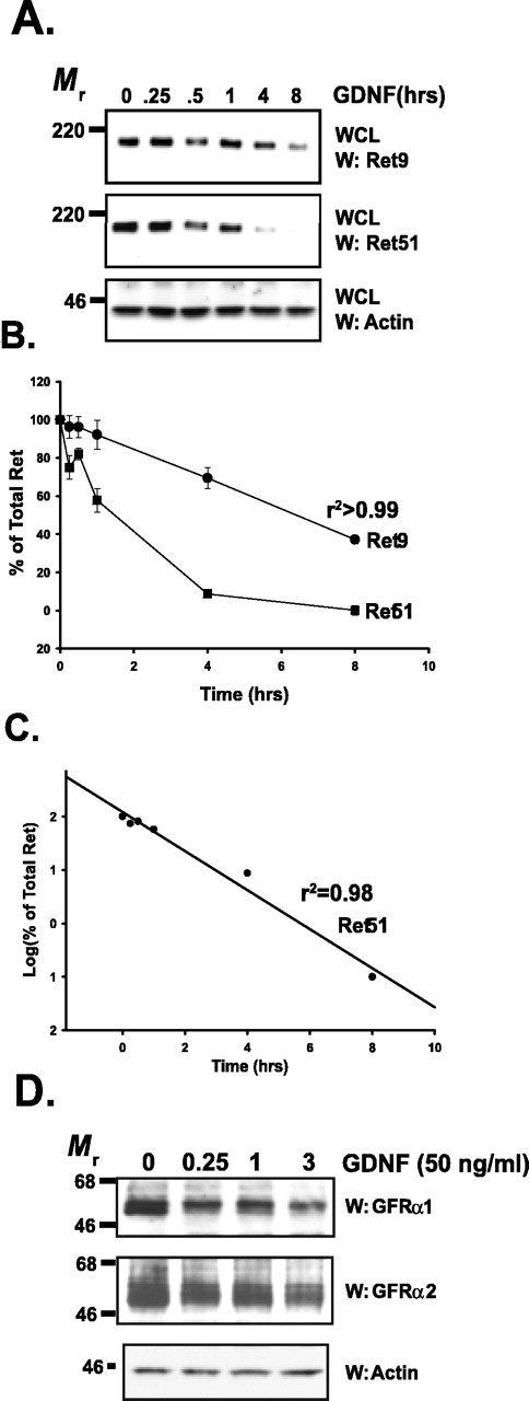
Figure 1.

Ret and GFRαs are degraded rapidly after activation. A, Sympathetic neurons were treated with GDNF (50 ng/ml) for various lengths of time, whole-cell extracts were produced from them, and the extracts were subjected to immunoblotting. Ret9 (top) and Ret51 (middle) immunoblotting revealed that both of these Ret isoforms are lost rapidly after GDNF stimulation. Actin immunoblotting (bottom) was used to confirm equal loading of cell extracts. WCL, Whole-cell lysate. B, Quantification of the experiment shown in A. Three separate experiments were conducted, quantified, and graphed as the mean ± SEM. C, Semilog plot of the data for Ret51 degradation kinetics shown in B. D, Sympathetic neurons were treated with GDNF (50 ng/ml) for various lengths of time (labeled on top), and whole-cell extracts were prepared. These extracts were immunoblotted with antibodies to GFRα1 (top) and GFRα2 (middle). Equal loading of proteins was confirmed with actin immunoblotting (bottom). This experiment was conducted twice with identical results. W, Western blot.
