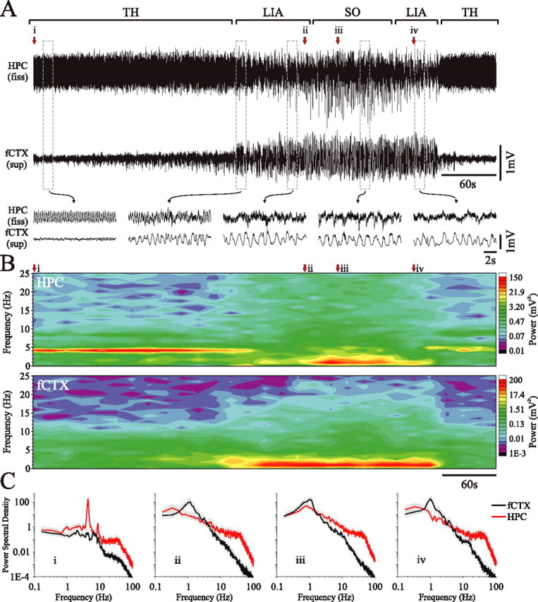
Figure 3.

Differential evolution of hippocampal and neocortical slow oscillatory states during urethane anesthesia. A, Simultaneous field recordings from the HPC and superficial frontal cortex during a spontaneous field state change. Bracketed segments (top) and expansions (bottom) of the traces highlight the features of the transitive evolution of the hippocampal SO compared with the neocortical SO. The beginning (and end) of the episode was characterized by activated patterns in both hippocampal (theta) and neocortical (LVFA) traces. The transition to slower and larger amplitude rhythms (culminating in the SO) appeared first in the nCTX, whereas the SO in the HPC appeared later, after an initial transition through LIA. This point is more clearly shown in the continuous spectrogram (B). C, Power spectra for fixed 60 s episodes beginning at points i, ii, iii, and iv as indicated in A and B (gray shaded area represents 95% confidence interval). TH, Theta; fiss, fissure.
