
Figure 6.
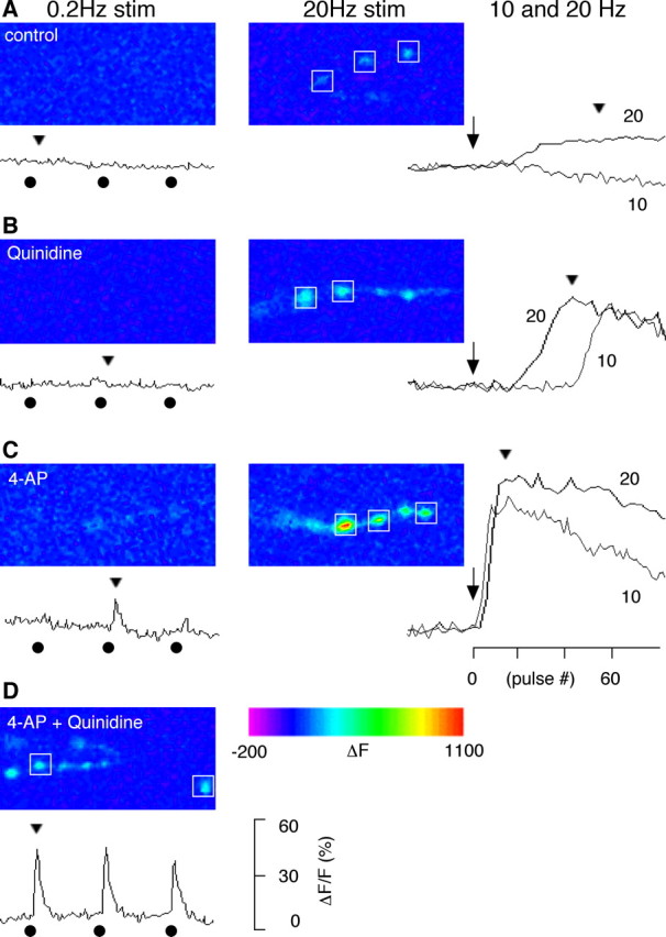
Altered Ca2+ dynamics in the presynaptic boutons treated with blockers of Sh and Shab channels. Presynaptic Ca2+ dynamics were monitored with a Ca2+-sensitive green fluorescent protein expressed in motorneurons (GCaMP; see Materials and Methods). A, In the absence of K+ channel blockers, fluorescent intensity in WT boutons did not change with single nerve stimulation (delivered at 0.2 Hz), but increased during high-frequency stimulation (detectable at 20 Hz but not at 10 Hz). This indicates that high-frequency nerve stimulation causes accumulation of cytosolic Ca2+. B, In the presence of the Shab channel blocker quinidine (30 μm), a sudden increase of fluorescence was observed during high-frequency stimulation (10 and 20 Hz). Note that the onset of the increase was faster for 20 Hz compared with 10 Hz. Single stimulation was not sufficient to trigger Ca2+ increase. C, In the presence of the Sh channel blocker 4-AP (200 μm), single nerve stimulation could sometimes trigger a small Ca2+ transient. However, the presynaptic Ca2+ accumulated very rapidly with high-frequency stimulation. D, When blockers for both Sh and Shab channels were applied, single nerve stimuli consistently triggered a fluorescent transient greater than those enhanced by 4-AP alone. Both the kinetics and amplitudes of fluorescent signals from individual boutons were similar within a nerve terminal branch, therefore, the sample traces shown are the average of signals from boutons indicated by a box (□). The onset of 10 and 20 Hz repetitive stimulation (arrows), the delivery of the 0.2 Hz test stimuli (•), and the time point for the displayed image frames (arrow heads) are indicated along the ΔF/F traces. The number of nerve terminal branches was 3–6 for each condition.