
Figure 1.
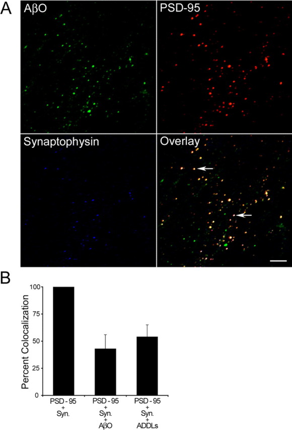
AβOs colocalize with synaptic markers in HCNs. A, Triple immunofluorescence of 18 DIV HCNs with AβOs (A11, green; 1:2500), anti-PSD-95 (red; 1:1000), and anti-synaptophysin (blue; 1:500). Cultures were incubated with 5 μm AβOs for 1 h before fixation. The overlay image shows the colocalization of the three antibodies. Triple colocalization is observed as light yellow fluorescent spots (arrows). Scale bar, 5 μm. B, Quantification of the frequency of colocalization of AβOs and ADDLs (antibody A11) with the synaptic markers PSD-95 and synaptophysin (Syn.). Error bars indicate the mean ± SEM.