
Figure 3.
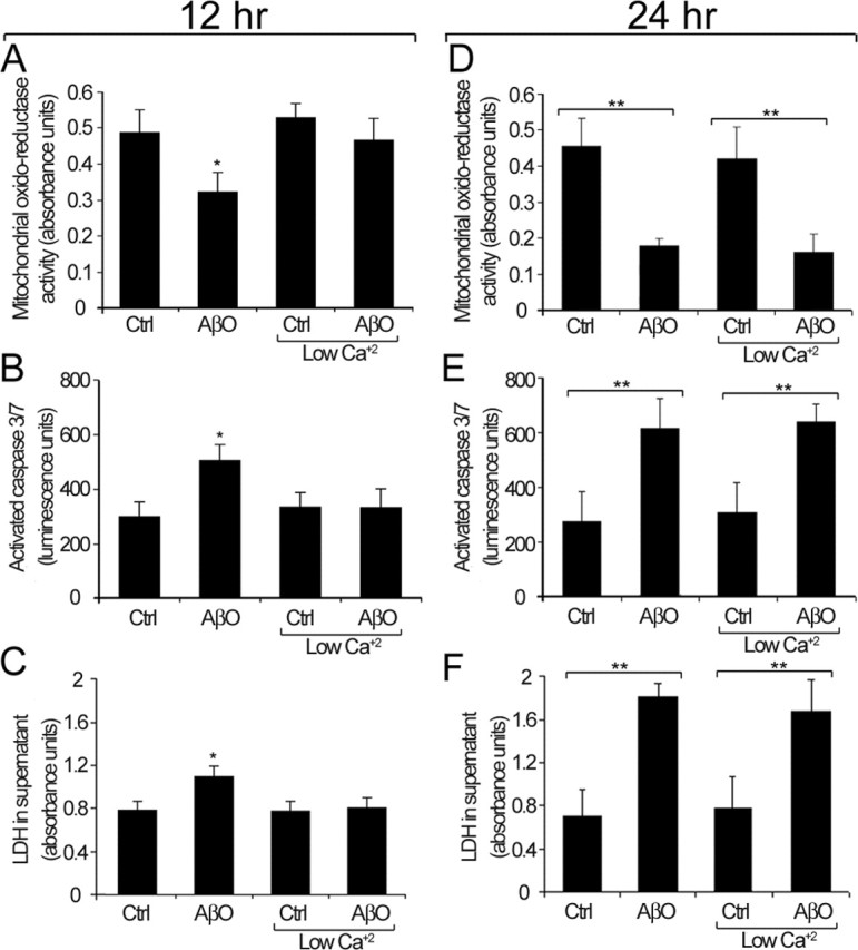
AβO toxic effect under normal and low extracellular calcium. A, D, Mitochondrial oxidoreductase activity at 12 h (A) and 24 h (D). B, E, Caspase 3 and 7 activation at 12 h (B) and 24 h (E). C, F, LDH levels in the culture medium at 12 h (C) and 24 h (F). Under normal culture conditions, these three parameters are significantly different from controls (Ctrl) after 12 h of AβO treatment (A–C). Under low calcium conditions, no significant differences in any of these parameters are observed between Ctrl and AβO-treated cultures at 12 h (A–C). By 24 h, there is a significant enhancement of neurotoxic parameters in AβO-treated cultures under both normal and low calcium conditions (D–F). Similar results were obtained in four independent experiments. Values are expressed as the mean ± SEM by unpaired Student's t test; ∗p < 0.05; ∗∗p < 0.01.