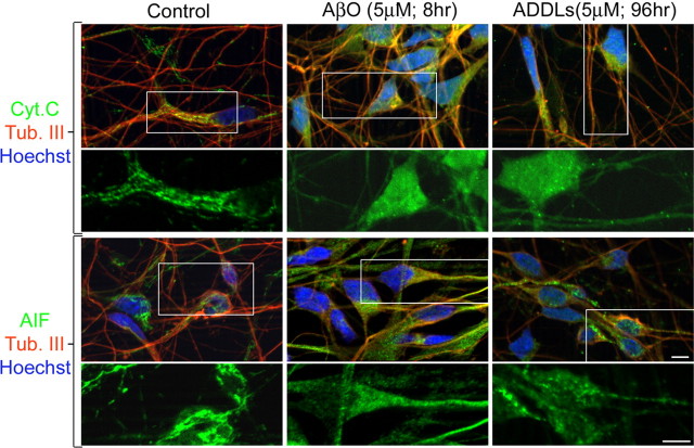
Figure 5.
Soluble AβOs induce translocation of cyt c and AIF from mitochondria to the cytoplasmic compartment. Shown is immunofluorescence of HCNs treated with vehicle (Control), AβOs, or ADDLs for the indicated times. Control cultures incubated with vehicle for 96 h exhibit mitochondrial localization of cyt c and AIF in cell bodies and processes (green fluorescence). In contrast, AβO- and ADDL-treated cultures exhibit diffuse cyt c and AIF localization in both cell bodies and neuronal processes (green fluorescence). The bottom panels are higher magnifications of the boxed areas. Scale bars, 10 μm. Neurons were double-labeled with the neuronal-specific marker anti-tubulin Class III (red fluorescence). Nuclei were counterstained with Hoechst (blue fluorescence).

