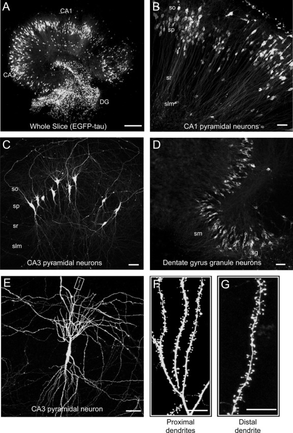
Figure 2.

Distribution of infected neurons in cultured hippocampal slices. A–D, Immunofluorescence micrographs of the whole slice (A), CA1 region (B), CA3 region (C), and DG region (D) of EGFP–tau-infected slices 3 d postinfection. Note efficient infection of neurons in all regions. E–G, High-power magnifications of a single CA3 pyramidal neuron (E) and sections of the proximal (F) and distal (G) dendrites. Note intense EGFP fluorescence in all cellular compartments. Slices were fixed and processed for immunofluorescence as described in Materials and Methods. so, Stratum oriens; sp, stratum pyramidale; sr, stratum radiatum; slm, stratum lacunosum-moleculare; sm, stratum moleculare; sg, stratum granulosum. Scale bars: A, 300 μm; B–E, 50 μm; F, G, 10 μm.
