
Figure 1.
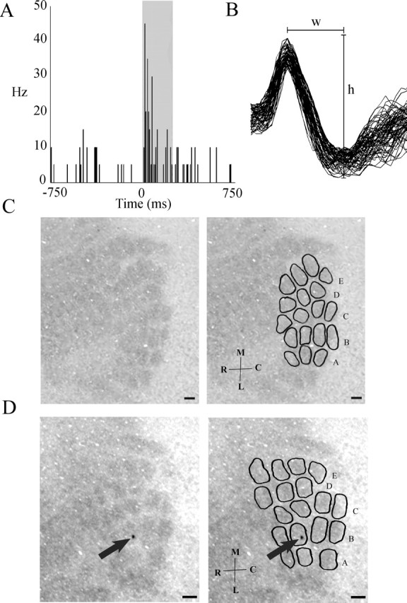
Cytochrome-oxidase-stained cortical barrel representations correspond to whiskers on the rabbit's face. A, Extracellular cortical single unit activity in response to 250 ms stimulation of B-row whiskers (gray box corresponds to the time of stimulation). Electrode coordinates from dura and cortical surface were 8.0 mm lateral, 2.75 mm posterior, and 1.3 mm ventral. One bin equals 10 ms. Note the increase in firing rate during whisker stimulation. B, Waveforms for the unit represented in A [width (w), 0.33 ms; height (h), 145 μV]. C, Left, Cytochrome-oxidase-stained rabbit barrel cortex. Note the barrel-like pattern. Right, Same image with barrels and rows delineated. D, Left, Unaltered cytochrome-oxidase-stained rabbit barrel cortex from which the neuron in A was recorded. Note the marking lesion in barrel B3 (arrow). Right, Same image with barrels and rows delineated. Scale bars, 250 μm. M, Medial; L, lateral; R, rostral; C, caudal.