
Figure 1.
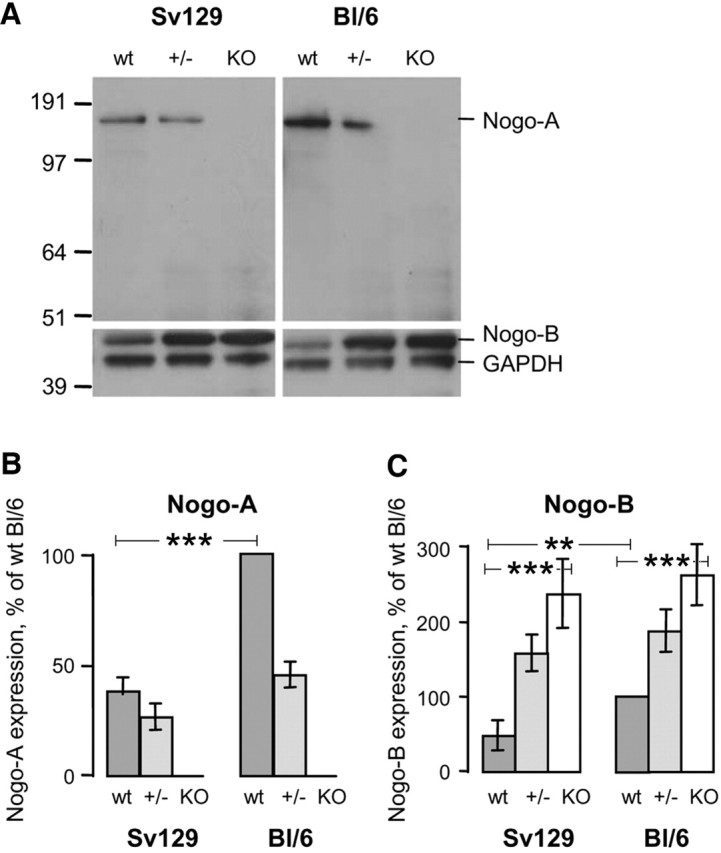
Endogenous Nogo-A and Nogo-B protein levels differ depending on the mouse strain. Levels of Nogo-A and -B in wild-type mice (wt) are higher in BL/6 than in Sv129 mice. In the homozygous Nogo-A knock-outs (KO), Nogo-A is absent and Nogo-B is upregulated three to five times in both strains. A, Immunoblotting with antiserum Bianca (Rb1) that recognizes Nogo-A and -B and an α-GAPDH antibody as internal standard. Total brain lysates from wild-type (wt), heterozygous (+/−), and homozygous Nogo-A knock-outs (KO) of 129X1/SvJ and C57BL/6 backgrounds are loaded in each lane. Molecular weight markers are indicated on the left. B, C, Densitometry of immunoblots for Nogo-A (B) and Nogo-B (C). Values are expressed in percentage of the BL/6 wild-type values.