
Figure 5.
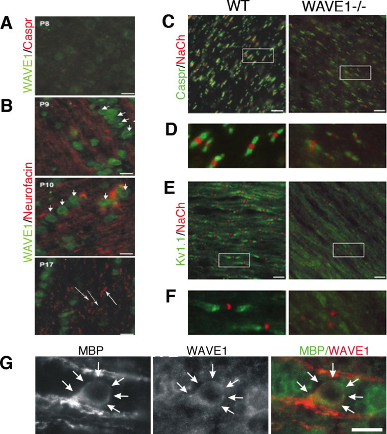
Clustering of Na+ and K+ channels at and near nodes is decreased in developing WAVE1−/− mouse optic nerve. A, Immunohistochemistry of P8 rat optic nerve revealed weak expression of WAVE1 and no expression of Caspr (red), a specific marker of paranodes. B, Increased expression of WAVE1 (green) is seen in P9 through P17 rat optic nerve. Expression of WAVE1 is mainly restricted to oligodendrocyte cytoplasm (short arrows). Neurofascin (red), a specific marker for nodes and paranodes, is evident in developing optic nerve (long arrows). C, P21 mouse optic nerve was double labeled with NaCh to mark the node and Caspr to mark the paranode and staining with NaCh and Kv1.1 to mark the juxtaparanode (E). Higher magnification of insets in C and E is seen in D and F. Nodal structure is normal in WAVE1−/−, but fewer nodes are seen. G, High-power image of an oligodendrocyte within the optic nerve double labeled for MBP and WAVE1. Scale bars, 10 μm.