
Figure 1.
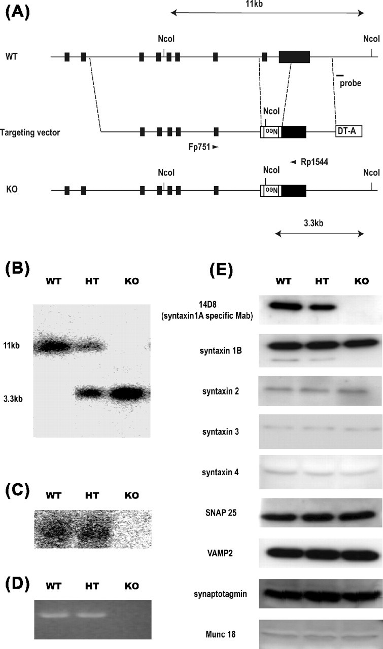
Generation of HPC-1/syntaxin 1A KO mice. A, Schematic showing the WT mouse HPC-1/syntaxin 1A locus, targeting vector construct, and targeted allele. The solid bar indicates HPC-1/syntaxin 1A exons 2–10. The targeting vector contains exons 4–10 of the gene. The region from exon 9 to exon 10 was replaced with a neomycin expression cassette (Neo). A diphtheria toxin expression cassette (DT-A) was attached to the 3′ end of the construct for negative selection. A probe for Southern blot analysis was indicated. B, Southern blot analysis of genomic DNA from a litter obtained by HT intercrossing. Each genomic DNA was digested by NcoI and hybridized with an external probe indicated in A. The 11 kbp WT and 3.3 kbp mutated DNA band were determined. C, Northern blot analysis of HPC-1/syntaxin 1A mRNA expression. Total RNA was prepared from postnatal day 7 mouse brain and hybridized with HPC-1/syntaxin 1A cDNA. The expression of HPC-1/syntaxin 1A mRNA was not detectable in KO mice. D, RT-PCR analysis of HPC-1/syntaxin 1A mRNA expression. A pair of primers, which corresponded to exon 8 and exon 10, was used. The expression of HPC-1/syntaxin 1A mRNA was completely lacking in KO mice. E, Western blot analysis of HPC-1/syntaxin 1A and other SNARE proteins. Whole-brain homogenate was prepared from postnatal day 7 mice. The expression of HPC-1/syntaxin 1A protein was lacking in KO, but other SNARE proteins were not affected.