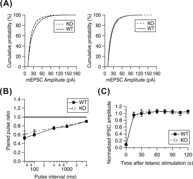
Figure 3.
Basal synaptic transmission in cultured hippocampal neurons from WT and KO mice. A, Cumulative histograms of mEPSC amplitude from individual events from KO or WT cultures at 7–8 and 14–15 DIV. At 7–8 DIV, the mean mEPSC amplitude of control cultures were larger than that of KO cultures (WT vs KO, 28.73 ± 0.53 vs 24.88 ± 0.46 pA), but at 14–15 DIV, no difference was observed (WT vs KO, 25.21 ± 0.34 vs 23.71 ± 0.17 pA; mean ± SE). B, Paired-pulse ratio of IPSC. There was no significant difference between cultured neurons from KO (n = 6) and WT (n = 12) mice. C, Recovery from depletion of synaptic vesicle by tetanic stimulation (20 Hz, 10 s). There was no significant difference between cultured neurons from KO (n = 3) and WT (n = 6) mice. Data represent the mean ± SE.

