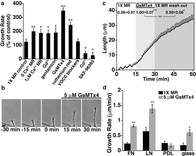
Figure 1.
Reducing Ca2+ influx through SACs increases neurite growth rates. a, Change in the average rate of neurite outgrowth during the 15 min period after various Ca2+ influx manipulations. Ca2+ influx was eliminated or reduced with 0 or low extracellular Ca2+ or blocked using Gd3+ (100 μm), gentamicin (200 μm), GsMTx4 (5 μm), ruthenium red (1 μm), a mixture of blockers for voltage-operated Ca2+ channels (1 μm ω-conotoxin, 100 nm nifedipine, 50 μm NiCl, and 60 nm ω-agatoxin), Cd2+ (50 μm), and SKF-96365 (3 μm). Each condition was normalized to the 15 min period preceding exposure to Ca2+ changes or blockers. Unless specified, each condition was presented in 1× MR solution (with 2 mm Ca2+ and 100 μm gentamicin). b, Representative images of an individual Xenopus spinal neuron axon at 15 min intervals before and after application of GsMTx4. Scale bar, 10 μm. c, The length of axons over a 60 min observation period before treatment (white shading), in the presence of 5 μm GsMTx4 (light shading), and after peptide washout (dark shading). For this graph, analysis was limited to responsive neurons (increased growth rate by at least 200%; n = 10 neurites). The average rates of outgrowth (micrometer per minute) during each period are shown above the corresponding graph. Similar results were found for all neurites tested (before treatment, 0.25 ± 0.04 μm/min; GsMTx4, 0.73 ± 0.05 μm/min; washout, 0.42 ± 0.05 μm/min; n = 67). d, Average rates of outgrowth 15 min before and after addition of GsMTx4 to neurons growing on different substrata. Blocking SACs with GsMTx4 significantly accelerates outgrowth on FN, LN, and glass but not on PDL. All averages are reported ±SEM. *p < 0.05; **p < 0.001. n ≥ 22 for all conditions.

