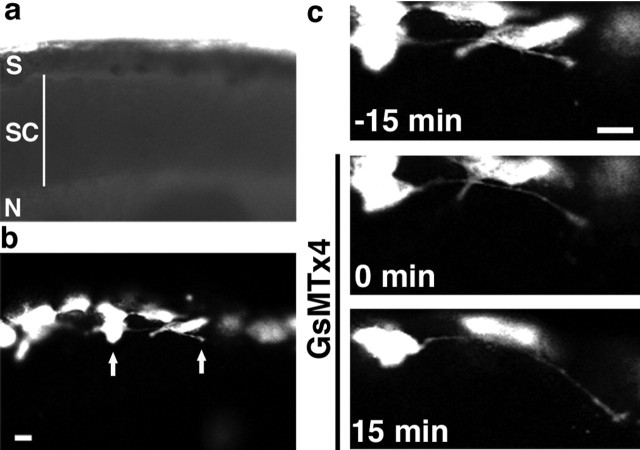
Figure 2.
Reducing Ca2+ influx through SACs with GsMTx4 increases neurite growth rates in vivo. a, Bright-field image of an embryo with skin and somites removed for in vivo confocal imaging. Dorsal is up, and anterior is to the left. S, Skin; SC, spinal cord; N, notochord. b, Fluorescence image of this embryo with GFP-labeled cells in the skin and dorsal spinal cord. Arrows indicate the cell body and growth cone of an extending axon. c, Time-lapse images of the labeled neuron in b at 15 min intervals before and after bath application of 5 μm GsMTx4. Scale bars, 20 μm.

