
Figure 6.
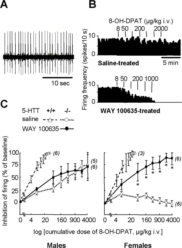
Functional status of 5-HT1A autoreceptors in the DRN. A, Oscilloscope trace showing spontaneous firing activity of a 5-HT neuron recorded in the DRN of a wild-type female mouse. B, Examples of integrated firing rate (in spikes per 10 s) histograms from female 5-HTT−/− mutants treated neonatally with saline (top) or WAY 100635 (bottom). Vertical bars above histograms represent injections of 8-OH-DPAT at the dose indicated (in μg/kg, i.v.). C, Dose-dependent inhibition by 8-OH-DPAT of DRN neuron firing in male (left) and female (right) 5-HTT−/− mutant mice after neonatal treatment with saline (open symbols, dotted lines) or WAY 100635 (filled symbols, solid lines). Inhibition of neuron firing (mean ± SEM of the number of neurons indicated in parentheses) is expressed as a percentage of baseline. Firing frequency was measured during the second minute after intravenous injection of cumulative doses of 8-OH-DPAT (on abscissa). The estrous cycle was not monitored because it does not influence this neuronal response (Bouali et al., 2003). *p < 0.05, significant difference for treatment (Fisher’s test). For reference data, 8-OH-DPAT-induced inhibition of neuronal firing was also assessed in groups of paired wild-type mice that had received neonatal treatment with saline (open triangles, dashed lines).